The financial and health-related impact of chronic wounds cannot be understated, as the cost of wound care has increased from £5.3 billion in 2012–13 to £8.3 billion in 2017–18. Furthermore, the UK is treating more patients — a rise of 1.6 million patients since 2012–13 with a small increase of 9% in a five-year period in healing rates (Guest et al, 2020). We can thus assume that the problem is getting worse and current provision for wound care is inadequate, although this does need to be balanced by the argument that we are potentially identifying more patients due to better assessment and documentation.  

Due to rising costs, there has been the development of the All-Party Parliamentary Group (APPG) on Vascular and Venous Disease, which has tried to highlight the extent of this problem in relation to venous disease and what can be done to improve care in the UK (APPG, 2019). The group has highlighted that the predominant interest of those involved in the management of vascular disease is targeted towards arterial disease, and ‘it is against this backdrop that venous disease sits in near silence’ (Carradice, 2020).  

The APPG also highlighted the inadequacy of the system with a failure of many GPs to diagnose and treat venous disease appropriately, with 75% of patients with known ulcers never receiving an appropriate assessment or being referred from primary to secondary care (Carradice, 2020). It was found that 64% of clinical commissioning policies were not compliant in providing access to venous treatment — in line with National Institute for Health and Care Excellence (NICE, 2013) guidance (APPG, 2019). This meant that over 90% of patients presenting with superficial reflux did not receive recommended evidence-based treatment within secondary care.  

Alongside this, in the author’s clinical opinion, it is vital to manage the long-term issues of chronic ulceration due to lymphovenous disease. Phlebolymphedema or lymphovenous oedema is the most common form of lymphoedema in the Western world — a secondary lymphoedema that develops in patients with chronic venous insufficiency (CVI) (Farrow, 2010). Contributing factors are increased survival of heart failure patients, medications associated with oedema, and increasing obesity (Fife and Carter, 2009; Farrow, 2010; Mortimer and Rockson, 2014; Todd, 2016). In the author’s clinical opinion, to improve outcomes for these patients, assessment skills need to be adapted to recognise lymphoedema, with treatment regimens considering oedema and not just assessing and treating patients purely for venous ulceration.  

BACKGROUND 


Mortimer and Rockson (2014) state that ‘the lymphatic system is fundamentally important to cardiovascular disease, infection and immunity, cancer, and probably obesity — the four major challenges in healthcare in the 21st century’. They demonstrated that lymphatic dysfunction is not a passive bystander, but an active player in relation to disease and should be targeted in relation to future therapeutic developments.  

Although lymphoedema is not directly responsible for leg ulcer development, it can affect wound healing (Mortimer and Browse, 2003). This is due to reduced oxygenation of the tissues resulting from the presence of oedema (Casley-Smith and Casley-Smith, 1997). As such, skin damage to an oedematous limb may lead to ulceration. Ulceration can also follow superficial infection (Green, 2007).  

End stage CVI leads to the development of lymphoedema, especially if there are recurrent periods of infection or inflammation due to the bacterial bioburden present in the wound bed. This prolonged oedema and lymphatic impairment can delay wound healing. This tends to be the case in most chronic, hard-to-heal leg ulcer patients seen by the author’s leg ulcer service, and it is these ulcers that need improved assessment and treatment to improve patient outcomes. 

The term chronic oedema acts as an umbrella description of abnormal swelling of tissues which lasts for more than three months, regardless of whether the aetiology is lymphatic or venous in origin (Humphreys et al, 2017) (Table 1). 
Table 1. Causes of chronic oedema (Lymphoedema Framework, 2006; Newton, 2011; Todd, 2016; Wound Care People, 2019) 
Overload Venous system malfunction leads to fluid overloading the lymphatics resulting in failure 
Insufficient lymphatics  Congenital abnormality can result in the absence of some lymph vessels from birth, or treatment of disease may require the surgical removal of lymph nodes 
Obstructed lymphatics  Lymph nodes or vessels can become obstructed by benign or cancerous growth 
Abnormal lymphatic contractability  The lymph vessels do not move fluid as well as they should 
Trauma to lymphatics  Damage may occur to the lymphatic system because of surgery or trauma 
Obesity Extra weight in the abdomen can put undue strain on the lymph vessels 
Immobility Puts undue strain on the lymph vessels 
Chronic venous hypertension  Resulting from failed or damaged valves in the leg veins, can lead to pooling of blood in the legs, resulting in oedema 
Mortimer and Rockson (2014) state that ‘it may be better to consider the presence of chronic oedema as synonymous with the presence of lymphoedema, as all oedema represents relative lymph drainage failure’. Peripheral oedema on assessment is usually classified according to possible systemic causes, such as local obstruction, heart failure, infection, nephrotic syndrome, injury, or tumours.  
 
The assessment approach taken needs to appreciate that there is usually more than one cause of oedema, taking into account the central role of the lymphatic system in drainage and tissue fluid balance (Mortimer and Rockson, 2014).  
 
The characteristics of chronic oedema are (Farrow, 2010; Bianchi et al, 2012; Ellis, 2015; Wound Care People, 2019):  
  • Swelling of more than three months’ duration, which remains on elevation 
  • Skin and tissue changes that may include: 
    • Dry, flaky skin 
    • Hyperkeratosis — hard,  scaly skin 
    • Skin creases, e.g. around the ankle and toes  
    • Fibrosis of the tissues 
    • Lymphangioma — blister-like bulging of dilated lymphatic vessels 
    • Papillomatosis — cobblestone effect on the skin due to lymphangioma and fibrosis 
    • Increased subcutaneous fat 
  • Tendency to bacterial and fungal infections 
  • Positive Stemmer’s sign (inability to pinch fold of skin at base of second toe due to thickening)

ASSESSMENT  

Thorough, holistic assessment is required to tailor care to the patient’s oedema type and the presenting problems within the wound bed (Wound Care People, 2019). This includes talking to the patient to understand their objectives and knowledge of their condition. In turn, this can help with treatment concordance if a good patient–nurse therapeutic relationship is established (Charles, 1995; Mandal, 2006; Morgan and Moffatt, 2008; Stanton et al, 2016). Indeed, compared to patients without lymphoedema, those with the condition present with poorer psychological adjustment and lowered health-related quality of life (Moffatt et al, 2017). 
 
A recent chronic oedema best practice document (Wound Care People, 2019) advocates a six-step assessment process:  
  • Story 
  • Self-care 
  • Site 
  • Skin 
  • Size 
  • Shape.  
This is a simple process to follow and can easily be incorporated into leg ulcer assessment and allows for more accurate assessment of oedema. 
Story
History and medical background
Ascertaining the duration of the oedema and when it started may help to indicate cause, e.g. if the swelling has followed recent surgical intervention or an insect bite, or if the patient has had recurrent venous ulcers. Arterial status should also be assessed. This should include either an ankle brachial or toe brachial pressure index, as well as clinical and physicial examination of the leg and foot (British Lymphology Society [BLS], 2019), e.g. is there any family history of venous disease, primary lymphoedema or lipoedema? Assess nutritional status, as protein deficiency leads to lower oncotic pressure (a form of pressure in the circulatory system which encourages water to cross the barrier of the capillaries and enter the circulatory system, however when its low, it goes into the interstitial space which can cause oedema), which can cause oedema.  
All co-existing morbidities or significant medical history should be identified and documented. The following have a significant effect on either wound healing or the development of lymphatic changes: 
  • Restricted mobility 
  • Obesity 
  • Diabetes 
  • Heart failure 
  • Dependency  
  • Chronic venous hypertension (lipodermatosclerosis) 
  • Hip/knee replacements  
  • Surgery, potentially involving damage to or loss of lymph nodes, i.e. hysterectomy, prostatectomy (Farrow, 2010; Newton, 2011; Humphreys et al, 2017; Wound Care People, 2019).  
Psychological status and socio-economic circumstances 
 
A best practice statement encourages the assessor to ask questions relating to how the patient is managing or coping with their condition, and any issues related to quality of life and assistance with activities of daily living (Wound Care People, 2019). Lifestyle choices which may contribute to swelling need to be investigated, e.g. sedentary lifestyle or obesity. Are there any work life or social circumstances affecting the condition due to prolonged periods of standing and sitting? This is important as it can influence the type of compression that patients require, which is usually stiffer with potentially higher levels of pressure.  
 
Psychosocial assessment to determine the impact of the swelling on the patient is important, as this can adversely affect body image. Evaluating the patient’s range of movement and dexterity will highlight any difficulty they might have in reaching their feet to allow washing or application of hosiery. Exaggerated skin folds provide the ideal environment for fungal infection. Often, hosiery gathers and tourniquets here and repeated attempts to pull hosiery up can damage the skin (Todd, 2014). Pain should also be assessed, as 50% of patients experience long-term pain in the limb(s) (Moffatt et al, 2003). Almost one-third of patients with chronic oedema experience cellulitis, of which 27% of these episodes require hospitalisation for intravenous (IV) antibiotic therapy (Moffatt et al, 2003). It is important that any previous infections are also recorded.  
Medications  
At assessment, if patients have lymphoedema present this can lead to diuretics being prescribed by GPs to deal with the identified swelling. However, diuretics remove none of the waste fluid, as it is a heavier molecule fluid than just water itself, and so use of diuretics for patients with lymphoedema will merely increase concentration of proteins and macromolecules in the interstitial space and speed the inflammatory process, leading to irreversible skin and soft tissue changes and increased risk of cellulitis (Raymond et al, 2017; Murdoch, 2020). Thus, healthcare professionals need to establish if diuretics are solely to treat lymphoedema or for other medical conditions, such as heart failure, hypertension etc, as if the former and not related to heart failure, renal failure, or hypertension, a discussion should be undertaken with the general practitioner to discontinue their use to prevent further deterioration in the wound.  

It is vital to assess all medications that the patient is taking, in case they cause or exacerbate oedema (Table 2).  
 
Table 2. Medications that may cause oedema (Todd, 2016)  
Calcium channel blockers (e.g. amlodipine, felodipine ,etc) 
Non-steroidal anti-inflammatory drugs (NSAIDs) (ibuprofen, naproxen, gabapentin)   
Hormones (e.g. oral contraceptives, oestrogen , testosterone and tamoxifen) 
Steroids, e.g. prednisolone 
Diabetes medication (thiazolidinediones), e.g. pioglitazone, rosiglitazone 

Self-care

Healthcare professionals should evaluate how prepared the patient is to be proactive in their care and take responsibility for their condition, as compression therapy may need to be introduced which they will have to manage, or techniques such as simple lymphatic drainage (SLD) and exercises (Wound Care People, 2019). Patient willingness to participate in their care can improve outcomes alongside the normal treatment regimens provided by clinicians.  
 
Site 
The following points are important to check in this part of the assessment process:  
  • Is the swelling acute or chronic? 
  • Assess the whole limb and the abdomen for oedema 
    • Is there bilateral or  unilateral swelling?  
    • Is swelling localised or  more generalised?  
    • Is there an inflammatory oedema due to allergies or contact dermatitis? 
Bilateral swelling can indicate heart failure, chronic venous insufficiency, or nephrotic syndrome (Wound Care People, 2019). Patients with heart failure may present with swelling in both legs that extends into the trunk, usually accompanied by a dry cough, breathlessness on exertion or when lying flat. However, renal and liver conditions can also result in bilateral swelling. Oedema in one leg can indicate deep vein thrombosis (DVT) if accompanied with pain, tenderness to touch and redness. Swelling in one leg can also indicate CVI or malignancy (Wound Care People, 2019).  
 
The location of lymphoedema gives clues to the possible underlying causes and informs where compression therapy should be applied (Wound Care People, 2019). 
 

Skin assessment 

In 2007, the Bonn Vein study found 100% of participants with active venous ulcers also had a positive Stemmer’s sign, indicating lymphoedema (Rabe et al, 2013). To assess for lymphoedema, a simple, manual test can be performed to identify early soft tissue changes indicative of lymphoedema and fibrosis. The Stemmer’s Test, the Bjork Bow Tie Test, which is an expanded version of Stemmer’s Test, and the pinch skin test (Wigg et al, 2016; Bjork, 2013; Bjork and Hettrick, 2018) can be performed anywhere on the body. The Stemmer’s sign is to try to pinch and lift a skinfold at the base of the second toe. If you can pinch and lift the skin, Stemmer’s sign is negative, if you cannot, the sign is positive.  
 
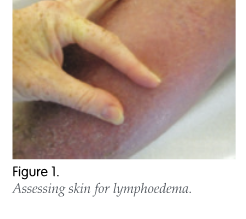
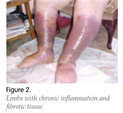
Skin that is positive for lymphoedema will be thickened, less pliable and produce limited or no ‘bow tie’ of wrinkles (Figure 1). This is indicative of chronic inflammation, tissue thickening and fibrotic soft tissue changes (Figure 2).  
Skin around a wound site should also be assessed by modifying the Stemmer’s to assess skin texture in affected areas (Bjork and Hettrick, 2018). This pinch skin test can be undertaken on the leg to detect areas of fibrosis and soft flowing lymphatics. It is important to feel the skin to see where the fibrotic changes end and there are ‘soft spots’, i.e. where the lymphatics are draining to (Wigg, 2016). These soft spots can be marked on a body map that identifies where wounds are. They can also be used to highlight areas of fibrotic, hard, or soft oedema.  
 
The following should also be assessed (Lymphoedema Framework, 2006): 
  • Sensitivities to  previous treatments  
  • Dry skin and hyperkeratosis (excess lymph fluid in the skin causes it to become thickened) 
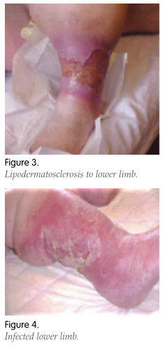
  • Signs of venous hypertension, haemosiderin staining, induration, varicose eczema or atrophy blanche, sub-malleolar venous flare, varicose veins and lipodermatosclerosis (Figure 3) (Keeley, 2009)  
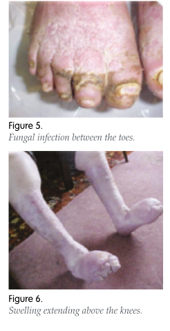
  • Any signs of infection or cellulitis (Figure 4). Lymphoedema means that the immune system is compromised, especially in the affected area. A delayed immune response and damaged lymphatics in the swollen tissue means that infection can occur suddenly. Cellulitis is nearly always unilateral (Opuku, 2015), so if redness and swelling is present in both legs, it is unlikely to be caused by cellulitis and a differential diagnosis should be considered, such as red legs or eczema 
  • Temperature and appearance of the surrounding skin (looking for peripheral arterial disease)  
  • Fungal infections, such as athlete’s foot, which commonly occur between toes and in skin folds (Figure 5) 
  • Lymphorrhoea — leakage of lymph through the skin. This occurs in untreated oedema and results in rapid swelling that the skin cannot accommodate. It appears as beads of fluid which put the affected area at risk of skin damage, since the skin becomes very wet and broken, increasing the likelihood of cellulitis (Morgan and Thomas, 2018) 
  • The wound bed should be assessed utilising a wound bed assessment tool, such as TIME (tissue, inflammation, moisture, edge of wound; Moore et al, 2019). However, the surrounding periwound area should be evaluated for fibrosis and any tissue hardening, as this will have a bearing on the microcirculation, oxygenation and nutrition of the wound bed (Williams, 2009).  
Shape 
Examine the shape of the limb: 
  • Is it regular or irregular? Does it resemble an inverted champagne bottle shape (Figure 3)? 
  • Are skin folds present? 
  • Is oedema pitting or non-pitting? 
  • Is swelling confined to the feet/foot or does it extend over the knees? (Figure 6) 
  • Are the toes affected? 
(Wound Care People, 2019) 
 
These factors will have a bearing on how the wounds and oedema are managed, effectively targeting compression therapy to oedematous areas and correcting the limb shape as far as possible. This may include adapting bandaging techniques to incorporate the toes, taking the compression therapy over the knee or to the thigh, introducing more complex techniques such as Kinesio® taping, the use of foam, and adapted bandaging techniques to break down fibrosis. 
 
 
Size 
 
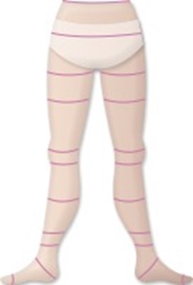
Limb circumference measurements, which evaluate the degree of swelling and monitor the effectiveness of treatment, should also be undertaken. Clinicians usually record calf and ankle measurements in uncomplicated venous disease, however, when lymphoedema is present, more in-depth measurements may need to be considered. Within lymphoedema clinics, measurements are taken at every 10cms from the floor to the thigh, although it may be easier to measure at the standard hosiery measurement sites (Figure 7).  
 
Comparison measurements are undertaken on a weekly basis by observing for a reduction in the circumference measurements. This helps to assess whether the compression is effective and when the oedema has potentially stabilised enough to measure for hosiery, as maintenance therapy when healed. If only one limb is affected, the other limb can be measured as a comparison (Lymphoedema Framework, 2006). 
 
Assessment should aim to identify what stage the oedema is, namely: 
  • Early stage — oedema usually presents as ‘pitting’ that reduces overnight or with elevation  
  • Disease progression — if allowed to progress untreated, the tissues become hard as waste products accumulate due to the lymphatic system’s inability to drain excess fluid, meaning that the oedema does not reduce with elevation or overnight  
  • Late stage — the affected tissues become hard, fibrosed and non-pitting; oedema does not reduce with elevation or rest and you begin to see pronounced limb-shape changes with evident skin folds and an increasing risk of episodes of cellulitis (Wounds UK, 2014).  

 
Figure 7. Standard compression hosiery measurement sites.

VASCULAR ASSESSMENT  

 
Vascular assessment should include either ankle brachial pressure index (ABPI) or toe brachial pressure index (TBPI) measurement. The ability to undertake a toe pressure should, where possible, be incorporated into the general assessment of all patients with lymphoedema, as ABPI readings can be elevated when oedema is present due to the volume of fluid present in the tissue, which can potentially lead to inaccurate readings giving a falsely higher ABPI (Doherty et al, 2006; European Society of Cardiology [ESC], 2011). Automated systems can be used, but clinicians need to ensure that they can interpret waveforms as well as the ABPI/TBPI reading.  
 
National guidelines recommend that vascular assessment be carried out as part of holistic assessment for patients with ulceration to exclude occult arterial disease before starting compression therapy for patients with leg ulceration (Clinical Resource Efficiency Support Team [CREST], 1998; Royal College of Nursing [RCN],1998; Scottish Intercollegiate Guidelines Network [SIGN], 1998). However, there is no such guidance for patients with lymphoedema (Wounds UK, 2009), resulting in many instances where ABPI/TBPI measurements are not undertaken due to the size of the limb.  
 
Cuff size is also important. Those suggested below are guidelines for arm circumference, but may also be applied to leg circumference:  
  • Limb circumference 22–26cm — small adult cuff 12–22cm 
  • Limb circumference 27–34cm — adult cuff 16x0cm  
  • Limb circumference 35–44cm — large adult cuff 16x36cm  
  • Limb circumference 45–52cm — adult thigh cuff 16x42cm  
(Wounds UK, 2009).  
 
Paediatric cuffs can be placed around the hallux to undertake toe pressures, but care needs to be taken to ensure that the readings are interpreted slightly differently, as a normal TBPI is above 0.7 rather than 0.8 in ABPI.
 
The size of the Doppler probe is important due to the signal distortion caused by the oedema in this patient group. It may be necessary to use a Doppler probe with a lower frequency than usual, a 5mHz rather than 8mHz, to locate the signal. 
 
Assessment of pedal pulses in patients with lymphoedema can be difficult due to the volume of fluid present in the tissue (Doherty et al, 2006). Although simple palpation techniques can be carried out, they are essentially flawed due to the distortion of the pulse signal through the oedematous tissue. 
 
Another useful resource when looking at vascular assessment is the utilisation of the British Lymphology Society (BLS) vascular assessment tool (BLS, 2019). This allows a simple checklist to be used to ascertain whether the arterial status is safe for the use of compression if you cannot perform a Doppler test.  

DISCUSSION  


A structural change within the NHS is needed to manage the increasing demand for wound care and improve patient outcomes (Guest et al, 2020). The author maintains that one of those changes is to train nurses in both tissue viability and lymphoedema to provide a one-stop service to patients, but also to educate general practice, community and district nurses to assess for the presence of lymphoedema, especially lymphovenous disease.  

Using a multidisciplinary team (MDT) approach is vital for venous leg ulcer management to promote continuity of care (Kjaer et al, 2005; Harding, 2006). However, the findings from the ‘Burden of Wounds’ study suggest that the MDT approach is not always available or implemented (Guest et al, 2016), impacting on patient care and outcomes. The National Wound Care Strategy Programme (NWCSP) recommends referral of patients to lymphoedema services, however this is dependent on availability and their willingness to treat patients with lower limb ulceration.  

A ‘seamless service’ through integration, coordination, and the sharing of information between different specialists and teams can promote continuity of care (Gulliford et al, 2006). The combined tissue viability and lymphoedema service at Healogics Wound Healing and Lymphoedema Centres offers this, which, in the author’s clinical experience, has led to better continuty of care, cost savings and improved outcomes

A best practice statement looking at holistic management of venous leg ulceration (Wounds UK, 2016) highlighted the presence and distribution of oedema that is more likely to become non-pitting with chronicity due to development of fibrotic tissue, but fails to mention that this is due to damage to the lymphatic system due to end-stage venous failure. However, a later best practice statement (Wounds UK, 2019) mentions that specialist bandaging techniques using inelastic compression may be required to accommodate unusual limb shape or to treat toe swelling. It also recommends referral to a lymphoedema service if available. In the author’s clinical opinion, a better approach would be for these patients to be seen by the same nurses and service. This would include changing the way we educate nurses in the community to include the assessment and treatment of lymphoedema at its earliest stages, rather than pure venous disease assessment and treatment.  

SUMMARY 

 
In the author’s clinical opinion, there are some simple changes that can be made to treatment regimens for patients with oedema, such as: 
  • Correctly assessing the cause and appearance of any oedema present 
  • Measuring the limb circumference regularly to monitor treatment  
  • Assessing for swelling to the toes and above the knee  
  • Knowing where soft spots are on the patient’s leg  
  • Assessing arterial status by utilising TBPI if able or using the BLS (2019) checklist.  
More complex patients with venous leg ulceration due to CVI and late-stage lymphoedema can be cared for by specialist tissue viability nurses with additional lymphoedema qualifications and training, reducing the requirement for two assessments and waiting for referrals to, in many cases, non-existent lymphoedema services.  
 
To effectively treat lymphovenous disease, it is vital to first assess the type of oedema that is presenting in order to treat the wound effectively. This requires being trained and competent in recognising lymphoedema, rather than concentrating purely on venous, arterial or mixed aetiology ulceration. The assessment process and documentation thus needs to adapt to reflect the changing needs of patients in relation to oedema.  

References

All Party Parliamentary Group (2019) Venous Leg Ulcers: A silent crisis. Available online: www.legclub.org/uploads/documents/b69cedd6ff0d355ce6c350db261af296.pdf  

Bianchi J, Vowden, K, Whittaker J (2012) Chronic odemea made easy. Wounds UK 8(2). Available online: www.wounduk.com 

Bjork R (2013) Positive Stemmer’s sign yields a definitive lymphedema diagnosis in 10 seconds or less. Wound Care Advisor 2(2):10–14 

Bjork R, Hettrick H (2018) Emerging paradigms integrating the lymphatic and integumentary systems: clinical implications. Wound Care Hyberbaric Med 9(2): 15–21 

British Lymphology Society (2019) Position Paper for Ankle Brachial Pressure Index (ABPI). Informing Decision-making before the Application of Compression Therapy. Available online: https://thebls.com/public/uploads/documents/document-20621539855354.pdf  

Carradice D (2020) Addressing the silent crisis of venous leg ulcers and its impact on patients. Available online: https://venousnews.com/addressing-the-silent-crisis-of-venous-leg-ulcers-and-its-impact-on-patients/  

Casley-Smith J, Casley-Smith JR (1997) Modern Treatment for Lymphoedema. 5th edn. Terrace Printing, Adelaide, Australia 

Charles H (1995) The impact of leg ulcers on patients’ quality of life. Prof Nurs 10(9): 571–3 

Clinical Resource Efficiency Support Team NI (1998) Guidelines for the Assessment and Management of Leg Ulceration.CREST Secretariat  

Doherty DC, Morgan PA, Moffatt CJ (2006) Role of hosiery in lower limb Lymphoedema. In: Lymphoedema Framework. Template for Practice: Compression Hosiery in Lymphoedema. MEP Ltd, London: 10–21 

Ellis M (2015) Lymphovenous disease and its effect on the lower limb. J Community Nurs 29(1): 32–40 

European Society of Cardiology (2011) ESC Guidelines on the diagnosis and treatment of peripheral artery diseases: Document covering atherosclerotic disease of extracranial carotid and vertebral, mesenteric, renal, upper and lower extremity arteries. Eur Heart J 32(22): 2851–2906  

Farrow W (2010) Phlebolymphedema: a common underdiagnosed and undertreated problem in the wound care clinic. J Am Col Certif Wound Spec 2(1): 14–23 

Fife C, Carter M (2009) Lymphoedema in bariatric patients. J Lymphoedema 4(2): 29–37 

Green T (2007) Leg ulcer management in patients with chronic oedema. Wound Essentials 2: 46–58 

Guest JF, Ayoub N, McIlwraith T, et al (2016) Health economic burden that different wound types impose on the UK’s National Health Service. Int Wound J 14(2): 322–30 

Guest JF, Fuller GW, Vowden P (2020) Cohort study evaluating the burden of wounds to the UK’s National Health Service in 2017/2018: update from 2012/2013. BMJ Open 10: e045253  

Gulliford M, Naithani S, Morgan M (2006) What is ‘continuity of care’? J Health Serv Res Policy 11(4): 248–50 

Harding KG (2006) Trends in wound care – the development of a specialty. Int Wound J 3: 147 

Humphreys J, Thomas M, Morgan K (2017) Managing chronic oedema in community settings. Wounds UK 13(3): 22-35  

Keeley V (2008) Drugs that may exacerbate and those used to treat lymphoedema. J Lymphoedema 3(1): 57–65 

Keeley V (2009) Chronic oedema of the lower limb: causes, presentation and management priorities. In: Skills for Practice: Management of Chronic Oedema in the Community. Wounds UK, Aberdeen 

Kjaer ML, Sorensen LT, Karlsmark T, Mainz J, Gottrup F (2005) Evaluation of the quality of venous leg ulcer care given in a multidisciplinary specialist centre. J Wound Care 14(4): 145–50 

Lymphoedema Framework (2006) Best Practice for the Management of Lymphoedema. International consensus. MEP, London  

Mandal A (2006) The concept of concordance and its relation to leg ulcer management. J Wound Care 18(8): 339–41 

Moffatt CJ, Franks PJ, Doherty DC, et al (2003) Lymphoedema: an underestimated health problem. QJM 96(10): 731–8 

Moffatt CJ, Aubeeluck A, Franks PJ, et al (2017) Psychological factors in chronic edema: a case-control study. Lymphat Res Biol 15(3): 252–61 

Moore Z, Dowsett C, Smith G, et al (2019)TIME CDST: an updated tool to address the current challenges in wound care. J Wound Care 28(3): 154–61 

Morgan A, Moffatt CJ (2008) Non-healing leg ulcers and the nurse–patient relationship. Part 1: the patient’s perspective. Int Wound J 5(2): 340–48 

Morgan K, Thomas L (2018) The development of a ‘wet leg’ pathway for chronic oedema. Int J Palliat Care 24(1): 40–6 

Mortimer P, Browse N (2003) The relationship between the Iymphatics and chronic venous disease. In: Browse N, Burnand KG, Mortimer PS. Diseases of the Lymphatics. Arnold, London: 293-6 

Mortimer PS, Rockson SG (2014) New developments in clinical aspects of lymphatic disease. J Clin Invest 124(3): 915–21  

Murdoch V (2020) Inappropriate use of diuretics and antibiotics for wet or ‘leaky’ legs. J Community Nurs 34(4): 58–62 

National Institute for Health and Care Excellence (2013) Varicose veins: diagnosis and managment. NICE, London. Available online: www.nice.org.uk/guidance/cg168 

Newton H (2011) Chronic oedema of the lower limb: pathophysiology and management. Br J Community Nurs 16(4): S4–12 

Opuku F (2015) Ten top tips: improving the diagnosis of cellulitis in the lower limb. Wounds Int 6(1): 4–9  

Rabe E, Hertel S, Bock E, et al (2013) Therapy with compression stockings in Germany — results from the Bonn Vein Studies. J Dtsch Dermatol Ges 11(3): 257–61  

Raymond R, Flanaghan M (2017) Chronic oedema: treatment and the impact of prescribed medicines. Pharmaceutical J online, 7 June. Available online: https://pharmaceutical-journal.com/article/ld/chronic-oedema-treatment-and-the-impact-of-prescribed-medicines 

Royal College of Nursing (1998) The Management of Patients with Venous Leg Ulcers. RCN, London 

Scottish Intercollegiate Guidelines Network (1998) The Care of Patients with Chronic Leg Ulcers. SIGN, Edinburgh  

Stanton J, Hickman A, Rouncivell D, et al (2016) Promoting patient concordance to support rapid leg ulcer healing. J Community Nurs 30(6): 28–35 

Todd M (2014) Strategies to prevent the progression of venous and lymphovenous disease. Br J Community Nurs 17(Sup9b): 3–14 

Todd M (2016) The challenges of caring for people with chronic oedema. Nurs Res Care 18(4): 198–200 

Wigg J (2016) Fluoroscopy guided manual lymphatic drainage from research to the classroom. International Lymphoedema Framework Conference/Australasian Lymphology Association Conference  

Williams A (2009) Chronic oedema in patients with CVI and ulceration of the lower limb. Br J Community Nurs 14(10): S4–8 

Wound Care People (2019) Best practice in the community. Chronic oedema. Wound Care People, Wixford. Available online: www.jcn.co.uk; www.gpnursing.com 

Wounds UK (2009) Skills for Practice: Management of Chronic Oedema in the Community. Wounds UK, Aberdeen  

Wounds UK (2014) Managing chroinc oedema with a venous leg ulcer — A quick guide. Wounds UK, London  

Wounds UK (2016) Holistic management of venous leg ulceration. Wounds UK, London. Available online: www.wounds-uk.com 

Wounds UK (2019) Best Practice Statement: Addressing complexities in the management of venous leg ulcers. Wounds UK, London. Available online: www.wounds-uk.com