

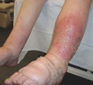
Figure 1. Cellulitis in a patient with lymphoedema.
TREATMENT FOR CELLULITIS OR ERYSIPELAS
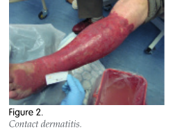
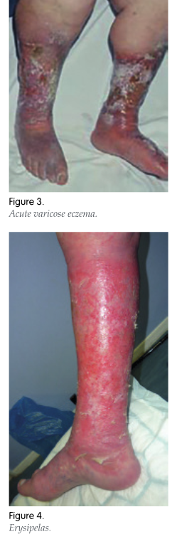
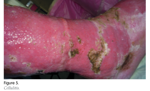
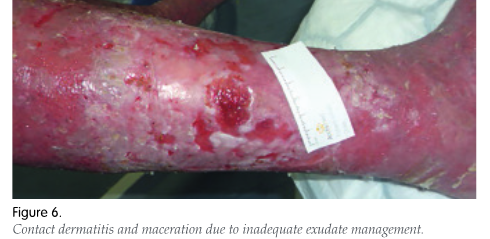
National Institute for Health and Care Excellence (NICE, 2019) guidance on managing cellulitis and erysipelas recommends excluding other causes of skin redness, such as an inflammatory reaction to an insect bite, or other conditions, such as chronic venous insufficiency, eczema or oedema. Furthermore, it recommends taking a microbiological swab only if the skin is broken and there is a penetrating injury, exposure to water-borne organisms, or if the infection was acquired outside the United Kingdom. Figures 2–5 show contact dermatitis, venous eczema, erysipelas and cellulitis.
It is recommended to draw around the extent of the redness with a single-use marker pen to monitor how it progresses before starting antibiotic therapy, but remember that the redness may be difficult to visualise in darker skin tones (NICE, 2019). People with cellulitis or erysipelas should then be offered antibiotics. However, healthcare professionals need to consider the site of the infection, severity of symptoms, risk of uncommon pathogens, for example from a penetrating injury, microbiological swab results and the patient’s meticillin-resistant Staphylococcus aureus (MRSA) status if known before prescribing
(NICE, 2019).
NICE guidance also recommends use of oral antibiotics as first-line treatment if the cellulitis and erysipelas are not severe and, in the case of intravenous (IV) antibiotics, to review the patient’s condition within 48 hours and revert to oral administration if there is evidence of improvement. NICE (2019) gives an extensive list of suitable antibiotics and practitioners are advised to consult their local trust formulary for guidance on which is the preferred type.
If symptoms worsen and the patient becomes systemically unwell, with increased pain out of proportion to the infection, or there is no sign of improvement after two to three days, it is recommended that healthcare professionals consider if there is another serious underlying condition, such as osteomyelitis, septic arthritis, necrotising fasciitis or sepsis (NICE, 2019). If a microbiological swab has not been taken, this should be done now, and the patient should be changed to a narrow spectrum antibiotic (NICE, 2019).
Santer et al (2018) note, however, that although cellulitis is usually treated with a one-week course of antibiotics, such as flucloxacillin, dependent on severity, comorbidity and site of infection, in many cases it does not resolve after one week and patients often receive repeated doses of antibiotics. They suggest that this may be unnecessary as the persisting redness can be due to inflammation, rather than active infection.
Santer et al (2020) also suggest that there is little evidence to guide the route of administration and that oral antibiotics appear to be just as effective as the IV route. As a result, they question the NICE Clinical Knowledge Summary guidelines, which recommend that patients with cellulitis, who are systemically unwell or who have diabetes, obesity, peripheral vascular disease, or chronic venous insufficiency, should be referred for either admission or IV antibiotics. Santer et al (2020) conclude that this advice is based on opinion rather than clinical evidence and that the majority of patients, with the exception of the systemically unwell, could be managed
at home.
This lack of guidance on prescribing antibiotics in cellulitis and erysipelas has also been highlighted by Bishop et al (2021) and Kilburn et al (2010). Beldon and Burton (2005) have produced a helpful algorithm for the management of limb cellulitis in primary and secondary care.
NICE (2019) does not recommend the routine use of prophylactic antibiotics to prevent future infections. However, the British Lymphology Society (BLS) guidelines recommend that patients with lymphoedema, who have had an attack of cellulitis, carry a two-week supply of antibiotics with them, particularly when away from home for any length of time, e.g. on holiday. Amoxicillin 500mg tds is recommended or, for those allergic to penicillin, erythromycin 500mg qds or clarithromycin 500mg bd (BLS, 2016).
Tissue viability management of ‘red legs’
As said, clinical signs of erysipelas/cellulitis can mimic other conditions, such as allergic contact dermatitis which is caused by exposure to an allergen or irritant substance that has damaged the normal barrier function of the skin (Beldon and Burton, 2005; Figure 6).
Clinically, these conditions will produce inflammation, oedema, pain, exudate, and blistering (English, 1997). In the case of venous disease, there may well be evidence of excessive keratin formation and thickening of the epidermis (Beldon and Burton, 2005). Patients with chronic venous leg ulcers may develop contact dermatitis as a result of long-term use of wound management products and the use of latex gloves (Tavadia et al, 2003). It is for this reason that latex gloves should be avoided.
If the patient has ‘wet’ cellulitis, venous eczema or contact dermatitis, the legs should be washed daily with a mild soap substitute, followed by moisturising with a bland emollient. Generic 50/50 liquid paraffin in soft white paraffin is the treatment of choice (Weller et al, 2015). Thirty minutes after applying an emollient, a potent steroid cream should be sparingly applied to the affected areas for a maximum of two weeks. If this does not resolve the skin’s condition, a reduced strength steroid cream can be used (Weller et al, 2015).
Exudate management may require the use of alginates or Hydrofibers as primary dressings, with absorbent secondary dressings, for example, Zetuvit® (Hartmann) or Exu-dry® (Smith and Nephew). However, in the initial phase, dressings may need frequent changing to prevent maceration (Beldon and Burton, 2005). Alternatively, superabsorbent dressings, such as Cutimed Sorbion Sachet Extra® (Essity) or Eclypse® (Advancis Medical), can be used. Healthcare professionals should consult their local trust’s dressing formulary for preferred choices.
If the patient is able to self-care, it may be more cost-effective to use a non-adherent contact layer, such as Adaptic™ (3M + KCI), Atrauman® (Hartmann) or Mepitel® (Mölnlycke), which can stay in place for several days, with the patient changing the outer dressing as required.
Potassium permanganate soaks are sometimes used as a weak antiseptic, however the effectiveness of these is debatable and there is no robust research evidence currently available to support their use. If using potassium permanganate, care must be taken to obtain the correct dilution of 1:10,000, as using a stronger solution may cause skin irritation (Beldon and Burton, 2005).
References
Atkin L (2017) Cellulitis of the lower limbs: Diagnosis and management. Nurse Prescribing 15: 588–92
Atzori L, Manunza F, Pau M (2013) New trends in cellulitis. WMJ Dermatology 1: 64–76
Beldon P, Burton F (2005) Clinical Practice Guidelines. Management guidelines for lower limb cellulitis. Wounds UK: 16-22.
Bishop J, Jones M, Farquharson J, Summerhayes K, Tucker R, Smith M, et al (2021) Implementation of a cellulitis management plan in three Australian regional health services to address an evidence–practice gap in antibiotic prescribing. Antibiotics 10(1288): 1–12
British Lymphology Society ( 2016) Consensus Document on the Management of Cellulitis in Lymphoedema. Available online: www.lymphoedema.org/wp-content/uploads/2020/01/cellulitis_consensus.pdf
Cheong HS; Chang Y, Joo E-J, Cho A, Ryu S (2019) Metabolic obesity phenotypes and risk of cellulitis: a cohort study. J Clin Med 8(7): 953
Cox NH, Colver GB, Paterson WD (1998) Management and morbidity of cellulitis of the leg. J R Soc Med 91(12): 634–7
Cranendonk DR, Lavrijsen APM, Prins JM, Wiersinga WJ (2017) Cellulitis: current insights into pathophysiology and clinical management. Neth J Med 75(9): 366–78
English JSC (1997) Contact dermatitis. Medicine 25: 42–5
El-Daher N, Magnussen CR (1996) Skin and soft tissue infections: outpatient management and indications for hospitalisation. Consultant 36(12): 2563–6
Eriksson B, Jorup-Rönström C, Karkkonen K, Sjöblom AC, Holm SE (1996) Erysipelas: Clinical and bacteriologic spectrum and serological aspects. Clin Infect Dis 23(5): 1091–8
Hofman D (1998) Oedema and the management of venous leg ulcers. J Wound Care 7(7): 345–8
Huttunen R, Syrjänen J (2013) Obesity and the risk and outcome of infection. Int J Obes 37: 333–40
Jorup-Rönström C, Britton S (1987)Recurrent erysipelas: Predisposing factors and costs of prophylaxis. Infection 15(2): 105–6
Kilburn SA, Featherstone P, Higgins B, Brindle R (2010) Interventions for cellulitis and erysipelas. Cochrane Database Syst Rev (6): CD004299
Kish TD, Chang MH, Fung HB (2010) Treatment of skin and soft tissue infections in the elderly: A review. Am J Geriatr Pharmacother 8: 485–513
Levell NJ, Wingfield CG, Garioch JJ (2011) Severe lower limb cellulitis is best diagnosed by dermatologists and managed with shared care between primary and secondary care. Br J Dermatol 164: 1326–8
Lipsky BA, Tabak YP, Johannes RS, Vo L, Hyde L, Weigelt JA (2010) Skin and soft tissue infections in hospitalised patients with diabetes: culture isolates and risk factors associated with mortality, length of stay and cost. Diabetologia 53: 914–23
National Institute for Health and Care Excellence (2019) Cellulitis and erysipelas: antimicrobial prescribing (NG141). NICE, London
National Institute for Health and Care Excellence (2020) Venous eczema and lipodermatosclerosis. NICE, London
Pavlotsky F, Amrani S, Trau H (2004) Recurrent erysipelas: risk factors. J Dtsch Dermatol Ges 2(2): 89–95
Pereira de Godoy JM, Galacini Massari P, Yoshino Rosinha M, Marinelli Brandao R, Foroni Casas AL (2010) Epidemiological data and comorbidities of 428 patients hospitalized with erysipelas. Angiology 61: 492–4
Santer M, Lalonde A, Francis NA , Smart P, Hooper J, Teasdale E , Del Mar C, et al (2018) Management of cellulitis: current practice and research questions. Br J Gen Pract 68(677): 595–6
Strazzula L, Cotliar J, Fox LP, et al (2015) Inpatient dermatology consultation aids diagnosis of cellulitis among hospitalized patients: A multi-institutional analysis. J Am Acad Dermatol 73: 70–5
Tavadia S, Bianchi J, Dawe RS, et al (2003) Allergic contact dermatitis in venous leg ulcer patients. Contact Dermatitis 48(5): 261–5
Weller R, Hunter H, Mann M (2015) Clinical Dermatology. 5th edn. Wiley Blackwell, West Sussex
Weng QY, Raff AB, Cohen JM, et al (2017) Costs and consequences associated with misdiagnosed lower extremity cellulitis. JAMA Dermatol 153(2): 141–6
Wingfield C (2009) Clinical review. Lower limb cellulitis: a dermatological perspective. Wounds UK 5(2): 26–36
Yosipovitch G, DeVore A, Dawn A (2007) Obesity and the skin: skin physiology and skin manifestations of obesity. J Am Acad Dermatol 56: 901–16